

Ready to be seen? Book a real appointment time online, 24/7 — Howell or Bloomfield Township. Takes about a minute, 24/7.
Medically reviewed by Dr. Tom Biernacki, DPM
Board-certified podiatric surgeon | Balance Foot & Ankle
Last reviewed: May 2026
The foot is one of the most neglected areas of the body in cancer surveillance — covered by socks and shoes most of the day, rarely examined during routine medical check-ups, and often dismissed as “just a wart” or “fungal” when something more concerning presents. In our Howell and Bloomfield Township podiatric clinics, we evaluate suspicious foot and toenail lesions regularly, and the cases that concern us most are the ones patients have been managing with OTC treatments for months before seeking care. The biology of foot cancer is no different from cancer elsewhere — early detection is the entire difference in outcomes.
Key takeaway: The most important rule in foot cancer recognition: the feet are checked last and least in dermatologic exams — patients rarely examine the soles and nail units themselves, and many physicians do not routinely remove shoes during check-ups. This means foot and toenail cancers are diagnosed at more advanced stages than equivalent lesions on the face or arms. Self-examination of the feet — including soles, between toes, and under nail edges — should be part of every monthly skin self-exam.
The statistic that changes how you think about foot cancer: subungual melanoma carries a 5-year survival rate below 30% for advanced cases — versus over 90% for early-detected cutaneous melanoma. The reason is a recurring diagnostic error: dark nail streaks are misidentified as fungus or bruising for 12–18 months while the tumor progresses to lymph node involvement. Every type of foot cancer covered here shares a pattern: it looks like something benign until it doesn't. Knowing which signs should never be dismissed can be the difference between a nail biopsy and an amputation. Call (810) 206-1402.
Subungual Melanoma: The Most Dangerous Toenail Cancer
Subungual melanoma arises from melanocytes in the nail matrix — the tissue that produces the nail. It accounts for 0.7–3.5% of all melanomas overall, but this proportion is significantly higher in people with darker skin tones: in African American, East Asian, and Hispanic patients, subungual melanoma represents up to 25–35% of all melanomas. The great toe and thumb are the most commonly affected digits.
Key takeaway: Subungual melanoma is the great masquerader. It is mistaken for a fungal nail infection, a bruise, a blood blister, and ‘just a dark spot’ for months to years before biopsy is performed. The two features that should immediately raise concern: a dark streak in the nail that was NOT caused by a specific remembered trauma, and any pigment extending from the nail onto the surrounding skin (Hutchinson’s sign).
Subungual melanoma typically begins as longitudinal melanonychia — a brown or black streak running from the nail base to the free edge. Over time, it widens, darkens, and develops irregular borders. The most alarming clinical sign is Hutchinson’s sign: extension of the pigmentation onto the nail fold (the skin at the base or sides of the nail). This finding is highly specific for melanoma and warrants urgent biopsy.
Advanced subungual melanoma causes nail destruction, nodular growth through the plate, ulceration, and bleeding. By this stage, regional and distant metastasis is common. 5-year survival: approximately 80% for localized disease; dropping dramatically with metastasis. Early diagnosis — when the lesion is still thin and confined — saves lives.
Diagnosis requires nail matrix biopsy. Dermoscopy can guide the index of suspicion — a parallel ridge pattern on the nail bed (the “parallel ridge pattern”) has the highest specificity for melanoma among dermoscopic features — but biopsy is the only definitive answer. We perform or coordinate nail matrix biopsies and have a low threshold for biopsy on any pigmented nail lesion without a clear trauma history.
A dark streak you cannot explain is worth an in-person look
Most pigment under a nail turns out to be a bruise or a harmless mole. The difficulty is that nobody can reliably tell which from a photograph, and this is a cancer that is very treatable when it is found early and much less so when it is not. The two features described above are the ones to act on: a streak you cannot trace to an injury you actually remember, and pigment spreading from the nail onto the skin around it. Either one is a reason to have the nail examined and, if there is any doubt at all, biopsied.
Howell — 4330 E Grand River Ave, Howell MI 48843 · Bloomfield Hills — 43494 Woodward Ave #208, Bloomfield Township MI 48302
Black Spot Under the Toenail: How to Tell a Bruise From Something Serious
This is the question that brings most people to this page, and there is one distinguishing feature that does more work than all the others combined: blood moves, pigment does not.
A subungual hematoma is blood trapped between the nail plate and the nail bed. It is attached to the underside of the plate, so as the nail grows out the mark travels forward with it and a band of normal, clear nail appears behind it. Melanoma is pigment being manufactured by cells sitting in the nail matrix at the base of the nail. The matrix stays where it is, so the pigment keeps being laid down in the same place. It does not march toward the tip and leave clear nail behind it. Over months it tends to persist, darken, or widen.
You can run this test at home. Mark the back edge of the discoloration with a fine pen, or photograph the nail next to a ruler in good light, and look again in six to eight weeks. Toenails grow slowly, roughly a millimetre a month, and a great toenail takes twelve to eighteen months to replace itself completely, so give it real time. A bruise should show visible forward movement within two to three months. Something that has not budged deserves an appointment rather than another round of waiting.
One caveat that matters more than any other on this page. A history of injury does not rule melanoma out. Most people can recall stubbing the toe or dropping something on it, because feet get knocked constantly. What frequently happens is that the injury simply draws attention to a mark that was already there. “I dropped something on it” is the single most common reason a subungual melanoma diagnosis gets delayed by a year or more. If the story does not fully fit, or the mark is not behaving like blood, it gets looked at regardless of what happened to the toe.
The ABCDEF rule for nail melanoma
Dermatologists use a purpose-built checklist for nails, because the familiar ABCDE rule for moles does not translate to a nail plate. It is a prompt for getting seen, not a scoring system that diagnoses anything:
- A — Age and ancestry. Peak incidence falls between the fifties and seventies. Acral melanoma makes up a substantially larger share of melanoma diagnoses in people of African, Asian, Hispanic and Native American ancestry.
- B — Band. Brown to black pigment, a breadth of 3 mm or more, and borders that are irregular or blurred rather than crisp and parallel.
- C — Change. A band that is widening, getting darker, or a nail problem that has not improved despite treatment aimed at something else.
- D — Digit. A single digit is more concerning than several. In the foot the great toe is by far the most common site.
- E — Extension. Pigment spreading out of the nail onto the surrounding skin fold or the fingertip.
- F — Family or personal history of melanoma or atypical moles.
Hutchinson’s sign, and the impostor that mimics it
Hutchinson’s sign is pigment that has spilled beyond the nail itself into the skin of the cuticle or the side folds. It is one of the more meaningful red flags in nail examination.
It also has a well-known look-alike. Pseudo-Hutchinson’s sign is pigment that is merely visible through a thin, translucent cuticle rather than actually present in the skin, and it shows up in several entirely benign situations. Telling the two apart depends on magnification and on viewing the nail from several angles, which is precisely why a photograph sent to a doctor cannot settle this one and an in-person look can.
The benign causes of a dark nail band that we see far more often
Perspective is warranted here. The overwhelming majority of pigmented bands we examine are not cancer:
- Ethnic melanonychia. Very common in people with darker skin tones. Typically several nails, bands of even width and uniform colour, present for years without change.
- Friction and pressure melanonychia. Caused by a shoe repeatedly loading the same nail. Classic in runners and hikers, and it favours the fourth and fifth toes and the great toe.
- Drug-induced pigmentation. Hydroxyurea, several chemotherapy agents, some antimalarials, minocycline and zidovudine can all darken nails. The tell is that multiple nails start changing at around the same time.
- A benign nail matrix nevus. Common in children, where a single band is far less worrying than a genuinely new single band appearing in an adult.
- Old subungual hematoma, which is the one that resolves itself on the grow-out test above.
What Happens if a Nail Lesion Needs Testing
People delay coming in because they imagine the visit ends in something drastic. It usually does not. The first step is simply a magnified, polarised look at the nail, which resolves a large share of cases on the spot. Under magnification the lines within a benign band are regular, parallel, and consistent in colour and spacing; the pattern that raises concern is irregular — lines varying in width, spacing and shade across the band, with loss of that parallel discipline.
If the appearance is genuinely concerning, the answer is a nail matrix biopsy. It is worth being straight about what that involves: the nail plate is lifted or a window is taken out of it, and a small sample is removed from the matrix under local anaesthetic. There is a real possibility of a permanent ridge or split in the nail afterwards. That is a fair trade, and patients almost always agree once it is framed honestly. A cosmetic nail defect is a small price for certainty about a melanoma.
The decision that actually costs people is not the biopsy — it is another six months of watching. An unexplained new band on a single digit in an adult is the specific scenario where waiting has the poorest track record. Acral melanoma is painless, and early on it looks unremarkable, which is exactly why it is so often found late rather than found small.
Why Melanoma on the Foot Is Found Later Than Melanoma Anywhere Else
Two reasons, and both are fixable.
The first is a widespread and understandable misconception. Because melanoma is so strongly associated with sun exposure, people assume the sole of the foot and the bed of a toenail are not places it can happen. Acral lentiginous melanoma — the subtype that occurs on palms, soles and under nails — is not driven by ultraviolet light. That is why careful sunscreen habits offer it no protection, and why it turns up at broadly similar rates across every skin tone. Melanoma being rarer overall in people with darker skin does not mean this particular form is.
The second is simply that nobody looks. The sole, the heel, the web spaces between the toes and the nail beds under polish are among the least-inspected square inches on the human body, and they are often not visible without deliberately picking the foot up.
The habit that fixes it takes under a minute a month. Sit down, put one foot across the opposite knee, and look at the sole, the heel, between every pair of toes, and all ten nails with the polish off. Ask whoever trims your nails to mention anything new or asymmetric. If you already come in for a diabetic foot exam, this inspection is part of it — an underappreciated benefit of an appointment most people think of as being only about circulation and sensation.
Anything new, changing, dark, or simply not healing on a foot is worth showing us, and we would far rather reassure ten people than miss one. There is no version of this where you have wasted our time.
Squamous Cell Carcinoma of the Foot
Squamous cell carcinoma (SCC) is the most common non-melanoma skin cancer of the foot and accounts for approximately 40% of foot skin malignancies. It arises from the keratinocytes of the epidermis and typically presents on sun-exposed dorsal foot skin — though it also occurs on the sole, between the toes, and periungualy (around the nails).
Key takeaway: Squamous cell carcinoma of the foot — typically arising from sun-exposed dorsal skin or from chronic wound/ulcer areas — may resemble a plantar wart, a callus, or a non-healing sore. The key red flag: any lesion on the foot that does not respond to standard treatment within 6–8 weeks and has been present for more than 3 months needs biopsy, not continued conservative management. If that describes your nail, book an urgent nail assessment online rather than waiting to see what it does next.
SCC on the foot most commonly appears as a rough, scaly, or crusted plaque; an ulcer with irregular raised edges; or a flesh-colored to reddish papule that does not heal. It is frequently mistaken for a plantar wart (verruca plantaris) — the key distinguishing feature: SCC bleeds with minimal manipulation, does not have the characteristic interrupted skin lines of a wart, and does not respond to standard wart treatments.
Risk factors for foot SCC: chronic sun exposure to the dorsum, history of radiation exposure, chronic non-healing wounds (Marjolin’s ulcer — SCC arising in the base of a chronic ulcer), human papillomavirus (HPV) subtypes 16 and 18 (associated with subungual and periungual SCC), immunosuppression, and prior arsenic exposure. Treatment: excision with adequate margins, Mohs micrographic surgery for periungual or cosmetically sensitive locations, and sentinel lymph node biopsy for high-risk variants.
Basal Cell Carcinoma
Basal cell carcinoma (BCC) is the most common skin cancer overall but is relatively rare on the foot (the foot receives less cumulative UV exposure than the face, scalp, and dorsal hands). When it does occur on the foot, BCC most commonly presents on the dorsal foot as a pearly, translucent papule with rolled borders and surface telangiectasias (tiny blood vessels). It rarely metastasizes but causes significant local tissue destruction if untreated. Treatment: excision, Mohs surgery for recurrent or aggressive subtypes.
Soft Tissue Sarcomas of the Foot
Soft tissue sarcomas — malignant tumors of connective tissue (fat, muscle, fibrous tissue, nerve sheath) — can arise anywhere in the body, including the foot and ankle. They account for approximately 5–10% of all foot tumors requiring biopsy in our experience. Common subtypes affecting the foot include synovial sarcoma (most common sarcoma distal to the knee), clear cell sarcoma (particularly associated with tendons and the foot), and epithelioid sarcoma.
Foot sarcomas typically present as a firm, deep, slowly enlarging mass — often painless in early stages, which delays diagnosis. The most important diagnostic clue: any soft tissue mass in the foot that is larger than 5 cm, deep to the fascia, firm, or enlarging needs MRI and multidisciplinary sarcoma team evaluation. Aspiration or surface biopsy without proper staging is contraindicated — it can seed tumor cells along the biopsy tract.
Kaposi’s Sarcoma
Kaposi’s sarcoma is a low-grade vascular tumor caused by human herpesvirus 8 (HHV-8), most commonly occurring in HIV-positive patients (epidemic KS) and in elderly men of Mediterranean or Eastern European descent (classic KS). The foot and ankle are among the most commonly affected locations — KS produces purple, red, or brown flat or raised nodular lesions on the skin, often starting on the feet and lower extremities. In HIV patients, KS may also involve visceral organs. The diagnosis is confirmed by skin biopsy.
⚠️ Foot and toenail lesions that require prompt biopsy or evaluation
- Dark streak or band in a toenail without a clear trauma history, especially if widening or irregular
- Pigment spreading from the nail onto the surrounding skin (Hutchinson’s sign) — high concern for subungual melanoma
- Non-healing ulcer, sore, or wound on any part of the foot present for more than 6–8 weeks
- Flat, scaly, red or brown patch on the foot that does not respond to antifungal or moisturizing treatment
- Fast-growing or deep lump or mass in the soft tissue of the foot or ankle
- Painful or bleeding lesion under the nail or on the sole that was assumed to be a wart but is not responding to wart treatments
Melanoma Without the Colour: Amelanotic Melanoma
Everything above assumes a dark spot. The most dangerous version on the foot is the one that is not dark at all, and it is dangerous precisely because it defeats every rule people are taught to look for.
Amelanotic melanoma produces little or no pigment. It appears as a pink, red, or plain skin-coloured bump, patch, or sore. There is no black spot, no brown streak, no irregular dark border — so it passes the ABCDE check and gets called something harmless. On the foot it is regularly treated for months as a wart, a callus that will not settle, a persistent ulcer, an ingrown nail, or a fungal infection before anyone biopsies it.
Because colour is unavailable as a clue, the useful signals are different ones:
- It is elevated. Raised above the surrounding skin rather than flat.
- It is firm. Solid to press, not soft or spongy like a blister or a cyst.
- It is growing. Steadily larger over weeks to months, without ever settling.
Those three — elevated, firm, growing — are the additional check used alongside ABCDE precisely to catch the lesions that have no colour to assess. Any one of them on a lesion that has been present for more than a couple of months is a reason to have it looked at properly.
Other features that should raise the question: a lesion that bleeds easily on light contact or catches on a sock; a small pink nodule with a slightly shiny or glassy surface; a lesion the same colour as your skin but with a subtle rim of pink around it; and anything that has been treated as a wart with no response at all.
The Rule That Catches What Colour Misses: Nothing on a Foot Takes Eight Weeks
If you remember one thing from this page, make it this: a lesion, sore, or lump on the foot that has not healed in about eight weeks needs to be examined and, if there is any doubt, biopsied. Not re-treated, not given another course of cream — examined.
Ordinary foot problems resolve. A blister heals in days. A bruise clears in weeks. A wart responds, at least partially, to treatment. A callus softens when the pressure is removed. When something on the foot simply keeps going, the reason is usually one of three things, and two of them matter:
- The pressure causing it has never been removed — genuinely common, and the reason a proper biomechanical assessment is part of the answer.
- It is not what it was labelled as — a “wart” that is a squamous cell carcinoma, a “callus” sitting over a melanoma, a “fungal” nail that is a subungual tumour.
- There is an underlying problem with healing — circulation, diabetes, or an autoimmune condition — which is itself worth identifying.
This rule matters more on the foot than anywhere else on the body, for reasons covered above: it is a site people do not examine, lesions are covered by socks and shoes all day, and everything down there has a convenient benign explanation ready to hand. Melanoma on the sole and under the nail is not rare because feet are protected — it is diagnosed late because feet are not looked at.
A biopsy of a foot lesion is a small procedure done under local anaesthetic in the office. Almost every one comes back benign, and that result is worth having. If you have a spot, sore, or nail change on your foot that has not resolved, have it examined rather than watched. Balance Foot & Ankle sees patients in Howell (4330 E Grand River Ave, Howell, MI 48843) and Bloomfield Township (43494 Woodward Ave #208, Bloomfield Township, MI 48302). Call (810) 206-1402.
Conditions Frequently Mistaken for Foot Cancer
Our most important diagnostic role is differentiating malignant from benign conditions — preventing both missed cancers and unnecessary patient anxiety. Common benign conditions that cause concern:
- Plantar warts (verruca plantaris): Cauliflower-like plantar lesion with interrupted skin lines, pinpoint bleeding (thrombosed capillaries) on debridement. Responds to wart treatments. Biopsy if atypical.
- Pyogenic granuloma: Bright red, rapidly growing, exophytic, easily bleeding lesion — benign vascular proliferation, not malignant. Can follow minor trauma. Treated by excision or cauterization.
- Glomus tumor: Benign vascular tumor under the toenail — produces exquisite point tenderness under one nail, worsens dramatically with cold. Characteristic triad: point tenderness, cold sensitivity, pain on nail pressure. MRI confirms. Treated by surgical excision through the nail.
- Clear cell hidradenoma: Benign adnexal (sweat gland) tumor — firm intradermal nodule, occasionally cystic. Biopsy required to confirm benign diagnosis.
- Subungual exostosis: Bony spur under the nail that lifts the nail and may bleed — not malignant but requires excision. X-ray confirms.
Having a Suspicious Spot on the Foot Examined
Almost everything that sends people to this page turns out to be benign. Most dark streaks in nails are bruises or normal pigmentation, most persistent sores are pressure or friction, and most lumps are cysts or fibromas. That is the accurate starting position, and it is worth holding onto.
The reason to have it looked at anyway is that skin cancers on the foot are found later than skin cancers anywhere else on the body, and that delay — not the cancer being more aggressive — is what drives the worse outcomes. Feet are not examined in the mirror, they are covered most of the day, and a spot on the sole or under a nail can grow for a long time without being noticed. An in-person look takes a few minutes and either resolves the worry or starts the process early, when it matters most.
It is worth being seen sooner rather than waiting if a nail streak is widening or the pigment extends onto the skin at the base of the nail, if a sore has not healed in a month or so, if a mole on the foot is changing, or if something bleeds or keeps recurring in the same place after apparently healing. Bring a photo if you have one from a few weeks or months ago — a comparison over time is genuinely useful information.
Balance Foot & Ankle sees patients at our Howell podiatry office and our Bloomfield Hills podiatry office. Call (810) 206-1402 or book as a new patient. If something needs a biopsy or a dermatology or oncology referral, we will arrange it rather than send you back to start again.
Frequently Asked Questions
How common is foot cancer? Foot and ankle cancers are uncommon relative to cancers at sun-exposed sites like the face and scalp, but they are consistently diagnosed at more advanced stages because of the foot’s relative obscurity in routine examination. Among all skin cancers, approximately 3–15% occur below the knee, depending on the study population. Subungual melanoma is the highest-stakes lesion to identify early.
Can a plantar wart turn into cancer? Ordinary plantar warts caused by low-risk HPV subtypes (types 1, 2, 4) do not become malignant. However, certain HPV subtypes (16, 18) are associated with a rare, malignant transformation of periungual and plantar tissue called Buschke-Löwenstein tumor (giant condyloma) or verrucous carcinoma — a low-grade variant of SCC that appears wart-like. Any wart-like lesion that is unusually large, has been present for years without responding to treatment, or has developed ulceration or unusual growth should be biopsied.
How do podiatrists check for foot cancer? In our clinics, we inspect the entire foot — including the sole, between all toes, and under and around the nail units — at every relevant visit. For any suspicious lesion, dermoscopy provides enhanced visualization of pigment patterns and vascular structures. For lesions that cannot be confidently diagnosed clinically, we perform or coordinate punch biopsy (for skin lesions), nail matrix biopsy (for nail unit lesions), or refer for MRI and oncology workup (for deep soft tissue masses).
The Bottom Line
Cancer of the foot, toes, and toenails is real, underdiagnosed, and — when caught early — highly treatable. The most dangerous lesson from foot oncology is the assumption that a dark toenail must be a bruise, or that a non-healing sore must be a wart. When in doubt, biopsy. The downside of a benign biopsy result is a small scar and reassurance. The downside of a missed subungual melanoma is measured in years of life.
Our Howell and Bloomfield Township teams evaluate suspicious foot and nail lesions regularly. We perform dermoscopic examination, coordinate nail matrix biopsies, and refer promptly to dermatology and oncology when indicated. Any lesion that concerns you deserves a professional opinion — same-day appointments are available.
Any Suspicious Foot or Toenail Lesion — Evaluated Same Day
Same-day appointments available in Howell & Bloomfield Township, MI
4.9★ | 1,123 Reviews | 3,000+ Surgeries Performed
Or call: (810) 206-1402
Sources
- Birch J, Thompson K, Goldenhersh M. Cutaneous malignancies of the foot. Clin Podiatr Med Surg. 2016;33(3):379-394.
- Levit EK, Kagen MH, Scher RK, et al. The ABC rule for clinical detection of subungual melanoma. J Am Acad Dermatol. 2000;42(2 Pt 1):269-274.
- Miettinen M, Fetsch JF. Evaluation of uncertain neoplasms in the foot. Clin Orthop Relat Res. 1997;(335):191-199.
- Nakamura Y, Ohara K, Kishi A, et al. Effects of non-surgical primary treatments on the local control of subungual melanoma. J Dermatol. 2012;39(9):782-787.
- Thai KE, Young R, Sinclair RD. Nail apparatus melanoma. Australas J Dermatol. 2001;42(2):71-83.
What are the most common types of cancer that affect the foot?
The most common malignancies affecting the foot are subungual melanoma (arising under the toenail), acral lentiginous melanoma (on the sole or heel), and squamous cell carcinoma. Soft tissue sarcomas can occur in the foot but are rare. Any growing, non-healing, or pigmented lesion on the foot or toe that does not resolve within 8 weeks should be evaluated and potentially biopsied.
Can foot cancer look like a fungal nail or a wart?
Yes — both subungual melanoma and squamous cell carcinoma can clinically mimic common benign conditions. A dark streak misidentified as a bruise, a scaly lesion mistaken for a wart, or a thickened discolored nail attributed to fungus may actually represent a malignancy. Any lesion that fails to respond to standard treatment, changes in morphology, bleeds spontaneously, or persists beyond 2 months warrants a biopsy.
How serious is subungual melanoma compared to other melanoma types?
Subungual melanoma carries a comparatively poor prognosis primarily because it is frequently diagnosed at a later stage — it is often mistaken for a fungal nail infection or traumatic bruise for months or years. Five-year survival rates improve substantially with early-stage diagnosis. Podiatrists and dermatologists are trained to recognize the clinical signs, making early evaluation critical if you notice a new or widening pigmented streak under a nail.
For a complete clinical overview: our detailed guide to nail melanoma warning signs — from a Michigan podiatrist — covering the Hutchinson sign, when to biopsy, and what to expect after diagnosis
📋 Dr. Tom Biernacki, DPM, FACFAS answers:
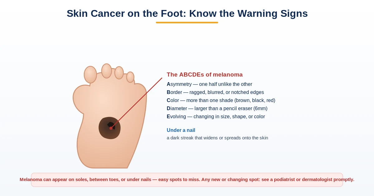
The most dangerous foot malignancy is acral lentiginous melanoma (ALM) — a form of melanoma that appears under or around the toenails, on the sole, or on the heel. It often starts as a dark streak under a toenail or a flat brown/black spot on the sole that people mistake for a bruise or age spot. The ABCDE rule applies: Asymmetry, irregular Border, multiple Colors (especially mixed brown, black, and pink), Diameter greater than 6mm, and Evolution (any change in size, shape, or color). A key ALM sign: a dark streak under a nail that extends to the skin at the base (Hutchinson’s sign). Any dark subungual pigmentation that doesn’t grow out with the nail, or any foot lesion that bleeds without injury, changes shape, or fails to heal in 4–6 weeks, needs biopsy. I biopsy any suspicious pigmented lesion — melanoma detected early is curable; detected late, it is not.
The American Academy of Dermatology emphasizes that any dark streak under a nail that changes in size, shape, or color should be evaluated promptly by a dermatologist or podiatrist.

Dr. Tom Biernacki, DPM is a board-certified foot & ankle surgeon (ABFAS & ABPM) at Balance Foot & Ankle Specialists in Southeast Michigan. With over a decade of clinical experience, he specializes in heel pain, bunions, diabetic foot care, sports injuries, and minimally invasive surgery. Dr. Biernacki is a member of the APMA and ACFAS, and his patient education content on MichiganFootDoctors.com and YouTube has made him one of the most-followed foot & ankle educators on YouTube.
More questions patients ask
Can you get cancer on your foot or toenail?
Yes — skin cancers including melanoma, squamous cell carcinoma, and basal cell carcinoma can occur on the foot. Subungual melanoma (under the toenail) is particularly serious and often mistaken for a bruise. Any dark streak, spot, or pigmented area under or around a toenail that doesn't resolve should be evaluated.
What does toenail melanoma look like?
Subungual melanoma often presents as a dark brown or black vertical stripe (melanonychia) running the length of the nail. Unlike a bruise, it doesn't grow out with the nail and may widen over time. Other signs include nail separation, bleeding, or pigmentation spreading to surrounding skin (Hutchinson's sign).
When should I see a doctor about a dark spot under my toenail?
See a podiatrist or dermatologist promptly if: the dark spot was not caused by a known injury, it doesn't grow out with the nail, it's widening, or pigment is spreading to the skin around the nail. Early detection of subungual melanoma dramatically improves outcomes.
What are signs of toenail cancer?
Concerning signs of toenail cancer (subungual melanoma): dark streak under nail extending into the cuticle (Hutchinson sign), uneven coloring, asymmetric borders, rapidly enlarging dark area, ulceration, bleeding, or splitting. ABCDEF rule guides assessment. Painless persistent unexplained pigmentation warrants biopsy.
Can you get cancer on your foot?
Yes. Foot cancers include subungual melanoma (under nails), acral lentiginous melanoma (palms/soles), squamous cell carcinoma, basal cell carcinoma, and rare sarcomas. Subungual melanoma is the most missed diagnosis — Bob Marley died from it. Any persistent unexplained foot lesion deserves dermatology or podiatry evaluation.
How is foot cancer diagnosed?
Foot cancer diagnosis requires biopsy (punch or excisional) of suspicious lesions. Dermoscopy aids initial assessment. MRI evaluates extent for confirmed cases. Sentinel lymph node biopsy stages melanoma. Time-to-diagnosis is critical: subungual melanoma 5-year survival drops dramatically with delay. Early biopsy of any concerning lesion is essential.
What is the most dangerous form of foot cancer?
Subungual melanoma — melanoma arising beneath the toenail or fingernail — is the most dangerous foot cancer due to its consistently late diagnosis. Because dark nail streaks are routinely attributed to fungal infection or trauma, the average time from symptom onset to correct diagnosis is often 12–18 months. By this time, the tumor has frequently advanced to Stage II or III. The 5-year survival rate drops dramatically with lymph node involvement compared to melanoma caught at Stage I.
What does foot cancer look like?
Foot cancer has several presentations depending on type: subungual melanoma appears as a dark brown or black longitudinal streak under the nail, often with Hutchinson's sign (pigment spreading to the surrounding nail fold). Squamous cell carcinoma of the foot appears as a persistent scaly patch, non-healing ulcer, or raised wart-like lesion that bleeds easily. Soft tissue sarcomas present as a painless deep mass or lump in the foot that grows slowly over months. Any non-healing lesion, new dark nail streak, or persistent growth on the foot warrants evaluation.
Is a dark spot under my toenail cancer?
Not necessarily — the most common cause of a dark area under the toenail is a subungual hematoma (blood pooling from trauma), which will grow out with the nail over 4–6 months. Toenail fungus can also cause dark discoloration. The concerning finding is a dark streak that does not grow out, widens over time, develops on a single nail with no trauma history, or is associated with Hutchinson's sign (pigment spreading onto the surrounding skin). A single evaluation with a podiatrist can distinguish between benign causes and findings that require biopsy.
What should I never ignore on my foot that could be cancer?
Six findings should never be dismissed without evaluation: (1) a dark longitudinal streak in a single nail that is new or widening; (2) pigment spreading from under the nail onto the surrounding skin (Hutchinson's sign); (3) a sore or ulcer on the foot that has not healed in 4 weeks; (4) a painless but slowly growing lump deep in the foot; (5) a scaly, red, or raised skin lesion that bleeds with minor contact; (6) any lesion that was previously stable but is now changing in size, shape, or color.
Still have a question about coverage or cost? Book online and we will check your benefits before your visit: Book in Howell · Book in Bloomfield Township. Prefer to talk it through first? Call (810) 206-1402.